§ CBCT Training

For institutions
In person · Online

Teaching clinicians to read the whole volume.

A structured programme in CBCT interpretation and reporting for dental schools, CPD providers and clinical groups. Taught by Dr. Shraddha Supnekar, MDS, Oral Medicine & Radiology, former Reader in Oral Medicine & Radiology at M.A. Rangoonwala College of Dental Sciences & Research Centre, Pune.

Start a conversation → Module outline
Audience
Dental schools, CPD providers, clinical groups
Format
In person or online
Duration
3-hour lecture series, or 3-day hands-on course
Cohort size
[PLACEHOLDER: participants per cohort]

§ 01 · Who it's for

Clinicians who prescribe, acquire or interpret CBCT.

01

Postgraduate trainees

Residents in oral medicine and radiology and allied specialties, working towards independent reporting.

02

Specialists and general dentists

Implant, endodontic, orthodontic and surgical clinicians who use CBCT in treatment planning.

03

Clinical teams

Dental groups with an in-house scanner that want a consistent approach to reading and reporting across the team.
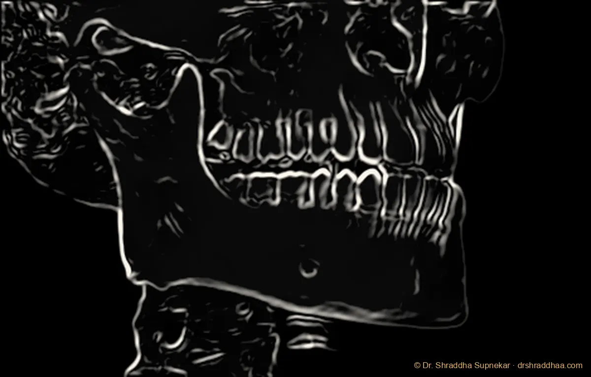
Line-drawing anatomy plate rendered from an anonymised CBCT volume, lateral view of the jaws and skull base, with the mandible, ramus, condyle, maxilla, zygomatic arch, occlusal plane, mental foramen and cervical vertebrae labelled Anatomical labels on the CBCT-rendered line drawingCONDYLERAMUSNASAL CAVITYMAXILLAOCCLUSAL PLANEMENTAL FORAMENMANDIBLE BODYCERVICAL VERTEBRAE
Anatomy plate rendered from an anonymised CBCT volume. Labelled: condyle, ramus, nasal cavity, maxilla, occlusal plane, mental foramen, mandible body, cervical vertebrae.

§ 02 · Modules

Two programmes.

01 · Undergraduates · general dentists

One-day lecture series · 3 hours

CBCT Fundamentals: When to Prescribe and How to Read

This course introduces the principles of CBCT, from basic physics and image reconstruction to radiation safety and dose comparison with other dental imaging. Technical aspects cover the components of a CBCT unit, field of view types, exposure factors, patient positioning, and common artifacts. Students learn normal CBCT anatomy and its applications in implantology, endodontics, oral surgery, orthodontics, and TMJ imaging. Emphasis is placed on when to prescribe CBCT using evidence-based selection criteria, along with its advantages, limitations, and the ethical responsibilities of the prescribing dentist.

02 · Postgraduates · maxillofacial radiologists

Three-day hands-on course

Mastering CBCT Reporting: Advanced Interpretation and Structured Report Writing

This advanced course builds on the principles of CBCT, covering cone beam geometry, voxel size and spatial resolution, reconstruction algorithms, and dose optimisation relative to 2D imaging and medical CT. Technical aspects address protocol selection, FOV and exposure optimisation, positioning errors, advanced artifact analysis, and multiplanar and curved reformatting for a systematic full-volume review. Clinical applications focus on interpreting complex cases across implant planning, endodontics, impactions, periodontal and orthodontic assessment, TMJ disorders, sinonasal and airway findings, and the radiographic differential diagnosis of cysts, tumours, fibro-osseous lesions, and systemic conditions. The course concludes with the advantages and limitations of CBCT and dedicated training in drafting structured, clinically relevant reports, including terminology, reporting of incidental findings, and medico-legal standards.

§ 03 · Outcomes

What participants can do afterwards.

Undergraduates · general dentists

  1. 01Explain the basic principles of CBCT and how it differs from 2D imaging and medical CT
  2. 02Decide when a CBCT is justified, using evidence-based selection criteria and ALARA/ALADA principles
  3. 03Choose an appropriate field of view for common clinical questions
  4. 04Navigate CBCT viewer software and scroll through axial, coronal, sagittal, and cross-sectional views
  5. 05Identify normal maxillofacial anatomy and key structures such as the inferior alveolar canal, mental foramen, and maxillary sinus
  6. 06Recognise common artifacts and avoid misreading them as pathology

Postgraduates · maxillofacial radiologists

  1. 01Optimise CBCT protocols, FOV, voxel size, and exposure parameters to balance diagnostic quality and dose
  2. 02Perform a systematic full-volume review using multiplanar and curved reformats
  3. 03Analyse complex artifacts and distinguish them from true findings
  4. 04Interpret complex cases across implantology, endodontics, impactions, orthodontics, periodontics, and TMJ disorders
  5. 05Evaluate sinonasal, airway, and other incidental findings beyond the dentoalveolar region
  6. 06Formulate radiographic differential diagnoses for cysts, tumours, fibro-osseous lesions, and systemic conditions
  7. 07Draft structured, clinically relevant CBCT reports with standardised terminology
  8. 08Communicate findings and recommendations clearly to referring clinicians
  9. 09Apply medico-legal standards in reporting, documentation, and handling incidental findings

§ 04 · Teaching background

Six years at the front of the room.

  • ▸Senior Lecturer (2019–2024) and Reader (2024–2025), Oral Medicine & Radiology
  • ▸Taught about 100 undergraduate students a year
  • ▸PG in-charge: co-guided seven postgraduate residents in CBCT reporting and thesis work
  • ▸Designed structured CBCT reporting modules for postgraduate trainees
  • ▸Conducted the department add-on course Basics of Cone Beam Computed Tomography (2023), 20 participants, interns
  • ▸MUHS university examinations (senior supervisor) and NAAC accreditation inspection
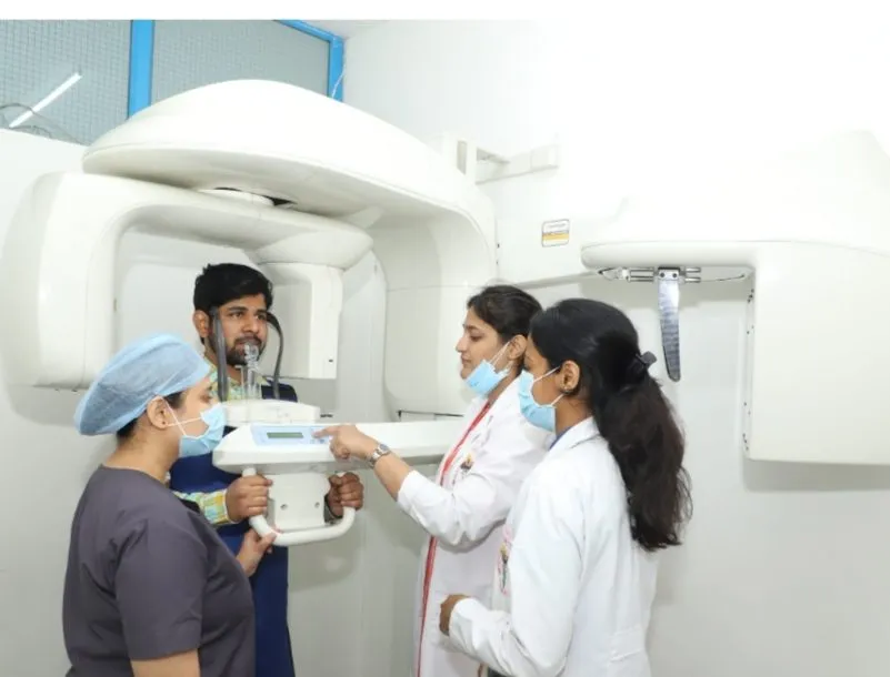
Demonstrating patient positioning in a panoramic and CBCT unit to dental students
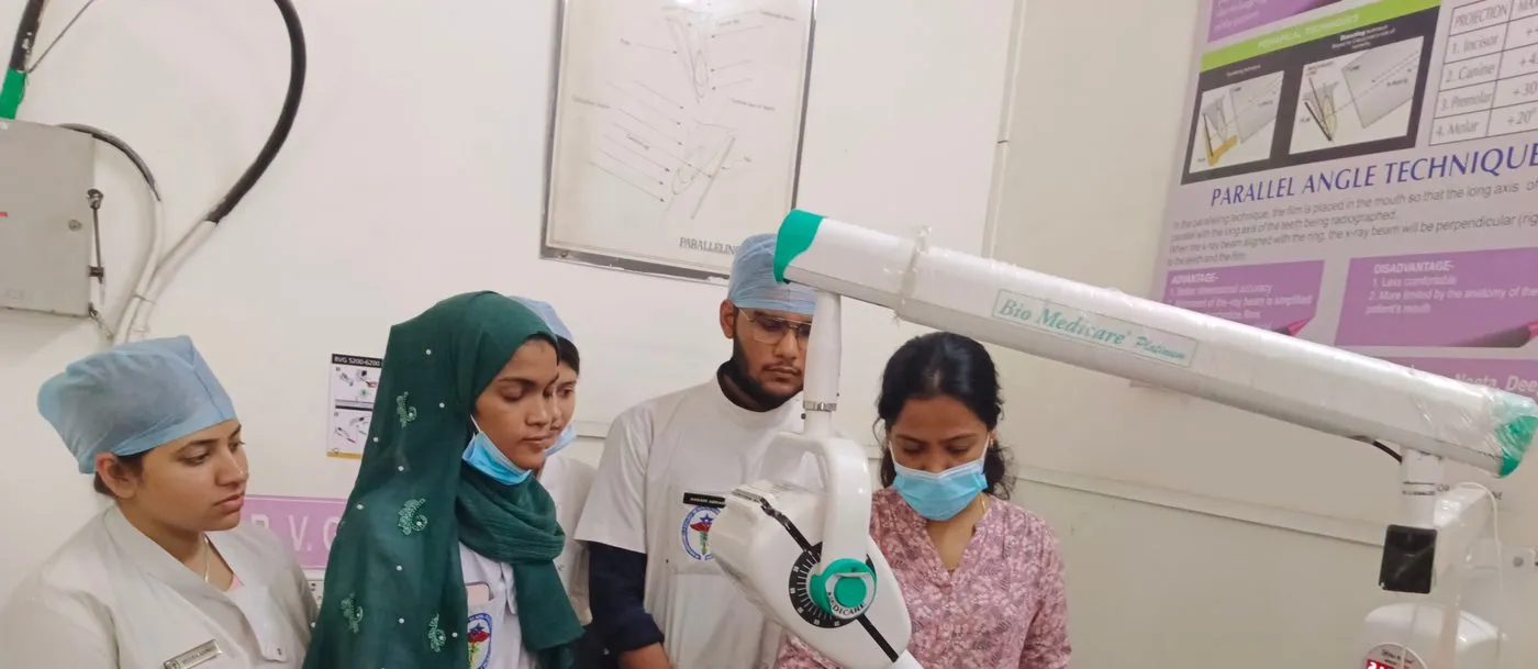
Dr. Shraddha Supnekar teaching intraoral radiographic technique on a phantom head to a group of students
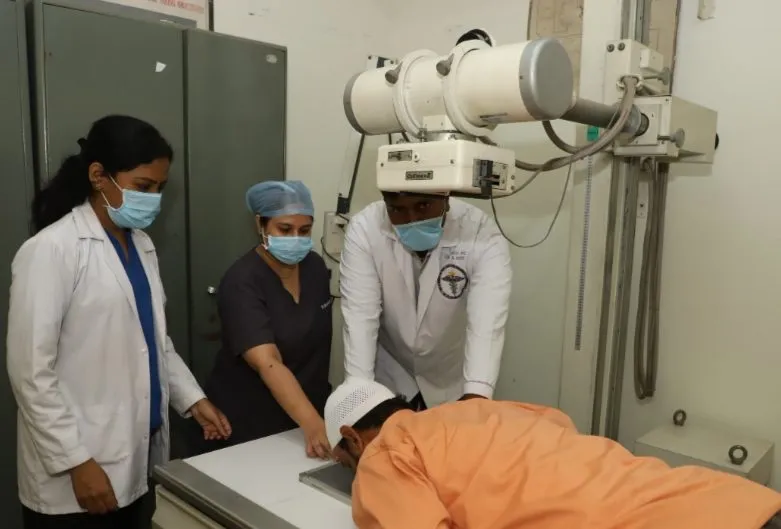
Demonstrating extraoral radiographic positioning to students
Radiography teaching · Department of Oral Medicine & Radiology, Pune

Planning a programme for your institution?

Share your audience, format and timing, and we can shape the programme around it.
Start a conversation → View CV